Monday, May 27, 2013

Using CBCT to Identify Vertical Root Fractures - Use of Filters

When we incorporated CBCT at Superstition Springs Endodontics, we hoped that it would be useful in identifying vertical root fractures.  There is a significant learning curve with the use of CBCT and identifying vertical root fractures may be one of the most difficult things to interpret.  The partnership with a good oral radiologist has also been an important part of learning to interpret CBCT.

In the past we have had to use clinical judgement, including presence of long, narrow periodontal pockets, j-shaped lesions as possible signs of vertical root fracture. However, those signs alone may also be caused by other conditions, not related to root fracture. Microscopic visualization was the most certain way to make this diagnosis. Too often, vertical root fracture is used as an excuse to remove a tooth without adequate evidence.

After more than two years of experience using high resolution, focus field CBCT in root fracture diagnosis, many consultations with the OMF Radiologist, we are more comfortable in diagnosing root fracture using CBCT, but the reality is that it is not always possible to see root fractures using CBCT.

Since the diagnosis of vertical root fracture (VRF) typically leads to extraction, it is our responsibility to be confident in that diagnosis.  In order to definitely see a root fracture using CBCT, it seems the fracture has to open up to some degree.  The more separation between fractured pieces, the better chance of making a definitive radiographic diagnosis. However, most vertical root fractures I see, do not have separated pieces.

Another challenge with identifying root fractures with CBCT is interpreting the difference between artifact (radiographic scatter) and actual fracture in the root.

Sometimes, a fracture cannot be seen with CBCT, but the bone loss pattern adjacent to a fracture can be seen. These bone loss patterns, not evident in traditional 2D imaging, can help identify the presence of a vertical root fracture.  Even with CBCT, all information has to be evaluated and weighed and a judgment to be made.  The only sure way is to see the fracture.  CBCT does allow us to "see" the fracture in certain instances but not all.

CASE REPORT


The following case would be typically diagnosed as a root fracture due to the bone loss pattern in the crestal area and into the furcation area as well as a long, narrow 8mm perio pocket on the ML surface.  There is however, coronal leakage under the mesial margin which could be a source of re-contamination of the root canal.


CBCT shows a long, periodontal breakdown on the lingual surface of the MB root - typical of a VRF.  Axial view also show the narrow bone loss in the same area - suggestive of a VRF.


Closer look at the axial view shows what appears to be a separation of the palatal side of the MB root.  This is a VRF - seen clearly on CBCT.


Rotation of the image, giving a palatal view of #14, shows a horizontal fracture in the MB root.

With these radiographic findings as well as the clinical findings, a confident diagnosis of vertical root fracture can be made.  Extraction was recommended in this case.


Wednesday, May 22, 2013

9.5 Year Recall of Apicoectomy

Last post, talked about how apicoectomies can be used to save teeth and the periodontium - tissues whose form and function are never truly replaced by dental implants. As a follow up to that post, here's a 9.5 year recall on an apicoectomy.


 This patient has crowns on #6-#11.  Tooth #6 is causing a localized ache, affected by pressure and chewing. #6 is sensitive to percussion with normal probings. DX:  Prior RCT with Symptomatic Apical Periodontitis (SAP). The margins are suspicious, but apical surgery was chosen to address the infection without disturbing the existing crown.

Apicoectomy completed, no root fractures seen with microscope. MTA retrofill.

9.5 year recall.  Tooth is fully functional, asymptomatic with radiographic healing.  Apicoectomy has preserved the tooth and the periodontium for an extended period of time.

Monday, May 20, 2013

Apicoectomies Save Teeth

Endodontic surgery has an established record of success.  Modern materials and equipment (microsurgery) make it even more successful.

In a recent updated meta-analysis of the literature regarding endodontic surgery (Tsesis et. al.), a successful outcome of endodontic surgery was seen in 89% of patients at 1 year.  It also pointed out that modern materials and equipment (MTA and microscopes) are associated with better outcomes.

The following case shows the benefit of endodontic surgery, use of CBCT in surgical planning, and the surgical correction of endodontic overfill.


This root canal was done by her general dentist 3 months earlier.  As you can see it is overfilled, post placed, crown prepped and ready for new crown, but patient continues to have symptoms with the tooth.


CBCT taken to evaluate the root morphology. It is clear that this is a single root/canal.  (The canal has an oblong shape)  It was chosen to treat this tooth with apical surgery to preserve the restorative work that has been completed and assure that the overextened gutta percha is removed.


Apicoectomy completed with MTA retrofill.


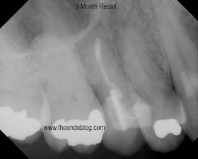
9 month recall show complete healing of the periapical lesion, tooth is fully functional and asymptomatic.  This procedure saves the patient significant time and money over extraction and implant placement.


SOURCES:

Outcomes of Surgical Endodontic Treatment Performed by a Modern Technique: An Updated Meta-analysis of the Literature
Igor Tsesis, Eyal Rosen, Silvio Taschieri, Yoel Telishevsky Strauss, Valentina Ceresoli, Massimo Del Fabbro

Journal of endodontics 1 March 2013 (volume 39 issue 3 Pages 332-339 DOI: 10.1016/j.joen.2012.11.044)