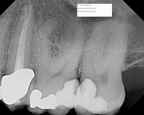
Dr. Hale's excellent post on pulp canal obliteration inspired me to share these few cases where a coronal barrier was also used to avoid root canal therapy. The most recognized reason to avoid complete pulpal debridement is biological, to maintain pulpal vitality, and thus continue root formation, subsequently improving fracture resistance, but there also exist technical limitations on the debridement procedure, imposed by anatomy or resorptive defects, that might prevent success of conventional root canal therapy.
This first example is a straightforward partial pulpotomy (or Cvek pulpotomy) with an MTA direct pulp cap. This patient had cerebral palsy and toppled out of his wheel chair causing a complicated (pulpal involvement) crown fracture of #10. You will note #9 was treated at this time as well, and if I recall correctly, was discolored and non-vital from a previous similar trauma. Multiple dental injuries (and traumatic injuries of all kinds) are very common in CP patients due to negative effects on balance. Fortunately, working with a pediatric dentist who scheduled OR time, the patient was seen within two days of the incident and the pulp vitality of #10 was maintained. Remember, inflammation in traumatic exposures very slowly spreads apically, and immature pulps with large vascular supplies are largely resistant to necrosis in the short term.
PreOp
Post-op

At a 1 year recall, #10 responded normally to vitality testing. Radiographs revealed a complete formed root and a dentin barrier beneath the MTA. Astute viewers will note this success is amazingly in the absence of a coronal restoration (unfortunately, not the only time I've seen bare, unrestored MTA pulp caps succeed at 1 year recalls).
This next case is similar, although a little less conventional. As you can see in the preoperative radiograph, the root is severely dilacerated. While certainly it is possible to perform root canal therapy on this type of root (see my previous post for an arguably more challenging S curve), the difficulty level is unquestionably high. This treatment plan not only reduces the risk of instrument separation, but also saves the patient time and money, and the operator from fatigue.
PreOp

Post-Op

The key here is that this was an asymptomatic carious pulp exposure. In the case of symptoms of irreversible pulpitis, it is generally thought that an MTA pulpotomy is a more risky procedure. It is certainly contraindicated in cases with symptomatic apical periodontitis (although I have had success direct pulp capping an immature tooth with apical periodontitis).
This last case is open to the most controversy. This patient had multiple large composite restorations across the anterior maxillary dentition. He admitted to being far more motivated by financials than esthetics. His previous composite restoration and crown had sheered off unconventionally at an oblique angle to the buccal leaving a substantial cingulum. The fractured portion had been rebonded by his general dentist. This tooth had a history of trauma over 40 years ago and some extensive external resorption is visible overlapping an obliterated pulp chamber and canal. The PDL is definitely in tact and there is no history of symptoms. The option of extraction and implant placement was discussed and encouraged. The alternative treatment plan chosen by the patient is less than ideal and the patient was more than okay with a compromised long-term prognosis. I intentionally described a grim outlook to the patient, as I do with most unconventional treatments, although here I can admit that I am confident in the predictability of the patient's choice. As you can see from the preop radiographs, conventional root canal therapy is impossible due to the irregular resorptive defect sandwiched between obliterated canal space.
PreOp
PostOp
I am still waiting on the general dentist to forward over a restored recall radiograph. Hopefully I will have the image to edit in by the end of the week. You can see the post space that I prepared using a 2 round bur and a gates-glidden with the tip flattened. The post space communicated with the resorptive more coronal than I anticipated, necessitating the use of MTA as a sort of resorptive cap. I feel as long as the area remains aseptic, it is reasonable to assume a successful result.
Here is a bonus case posted on our facebook page, http://www.facebook.com/pages/Alpharetta-Endodontics/137382942943581 . I'd encourage everyone to follow there (and check the backlog of case photos) for more interesting cases.
The patient's symptoms were intermittent, spontaneous, a 6 or 7 out of 10 on the pain scale, occasionally throbbing, and worse with mastication and pressure. The key history here is the patient's remark, "it feels like my gums are coming loose from my tooth."
Make the diagnosis. I have obviously helped by circling the key components.


Only a second year Dental Student here so who knows if my dx will even be close but I will give it a shot based on my current education...
ReplyDeleteThat white lacey looking lesion on the pt's buccal mucosa jumps out at me as lichen planus. Because the pt is feeling as if his/her "gums are coming loose from my tooth," the lichen planus would be considered erosive. Lastly, I am confident that blog post would have mentioned if these lacey white lesions were bilateral, but, because this was not mentioned, I will make the leap to say that it is only located where it is shown in the photographs. With this information, I would make the clinical diagnosis as erosive lichen planus.
In terms of treatment, after attempting to remove sources of irritation and xerostomia, possibly a topical corticosteriod like fluocinoide.
www.DentalDadDiary.com
Thanks for sharing this post!!!!
ReplyDeleteReally informative photos and x rays
ReplyDeleteThanks for the kind responses.
ReplyDeleteDental Daddy: Erosive Lichen Planus is indeed correct. Nice blog! Those denture wax-ups bring back some memories I'd rather keep forgotten :)
This is one of the well-described blogs on root canal. Very clearly explained... thanks for sharing!
ReplyDeleteThis is a very informative and helpful post. I look forward to reading more of the information you provide. Thank you.
ReplyDeleteThis is a great innovation. I think more people will opt for this procedure because having root canals discolors the teeth.
ReplyDeleteNice post regarding the problem faced by most of us related to root canals, and this post is very helpful
ReplyDeleteThis is an excellent post! Thank you for taking the time out of your busy dental schedule to share it.
ReplyDelete